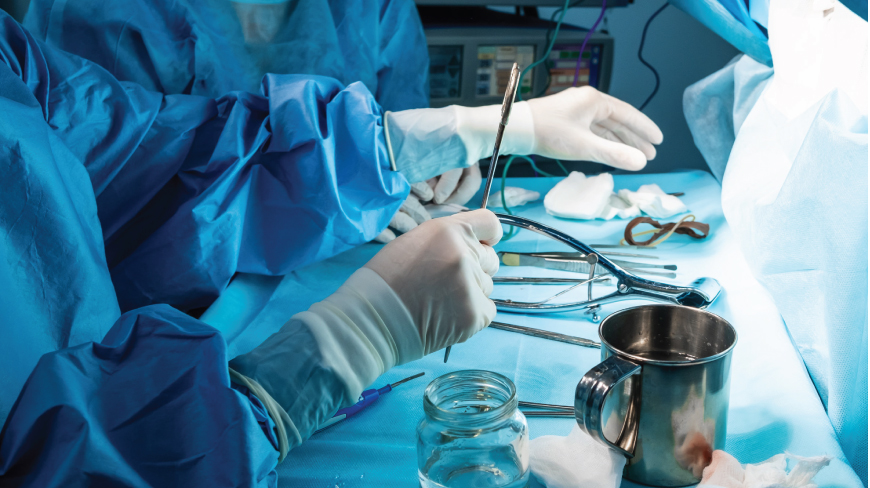
Fistula Surgery
Anal fistula is a chronic abnormal communication between the epithelialised surface of the anal canal and the perianal skin. There is a chance of 2 per 10,000 population per year to be affected by anal fistula. With the frequent peak incidence being higher between 30 to 50 years of age. Men are likely to be affected by this condition more than women. Anal fistulas do not heal themselves and surgery is the only curative modality available. Fistulas can significantly affect quality of life.
Techniques for fistula surgery
- The patient is placed in a prone jackknife position and the buttocks are parted for visualisation.
- Examination of the area is carried out under anaesthesia to determine the extent of fistula.
- Fistulotomy is a laying-open technique and is most useful for primary fistulas (submucosal, low transsphincteric and intersphincteric). A probe is inserted through the external and internal openings.
- Electrocautery is used to divide the internal sphincter, subcutaneous tissue and the overlying skin. The tract is opened in its entirety
- The wound is opened out about 1 – 2 cm adjacent to the external opening. The skin is locally excised to aid internal healing.
The procedure is carried out either in a stand-alone mode or in combination with a fistulotomy. Complex fistulas, immunosuppressed patients, multiple fistulas, anterior fistulas in women are best treated with a Seton placement.
Patients with chronic fistula are recommended the mucosal advancement flap. A total fistulectomy is carried out removing both the primary and secondary tracts inclusive of the internal opening.
Called the ligation of the intersphincteric fistula tract, LIFT is a procedure that is performed on complex transsphincteric fistulas. Preservation of the sphincteric muscles is a major feature of the surgery. The intershpincteric plane is accessed and the internal opening is secured with the removal of the infected cryptoglandular tissue.
